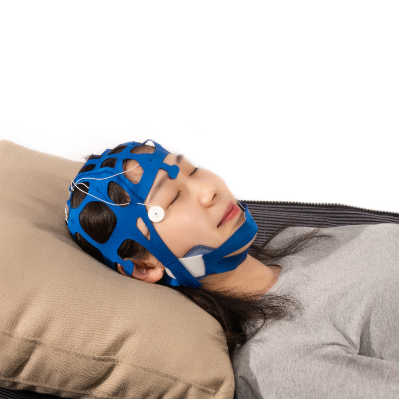
The product is used for fixation between human body and electrode wire
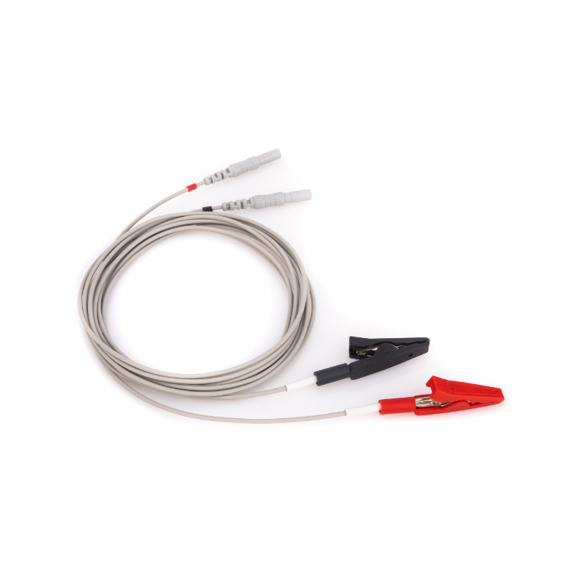
EEG (EMG) lead wire
【 Main structure】
Composed of instrument plug, signal wire (shielded and unshielded), metal electrode parts (metal pin, disk, clip, button).
【 Product performance】
1. Applied 10Hz sine wave and not exceed 100μA (p-p) current to electrode pair, The AC impedance of the electrode pair does not exceed 3kΩ.
2. After 1 min stable period, the DC offset voltage of the electrode pair is not more than 30mV.
3. The DC impedance at both ends of the wire is 0.5Ω-2.5Ω (for 1.5 m, when the length of the wire increases, the corresponding resistance increase should not exceed 1Ω/m).
【 Application】
Used with a variety of EEG, EEG Sleep monitors, EEG analyzers, and EMG analysis systems to acquiring and transmit human bioelectric signals.
【 Contraindications】
It is forbidden to use this product if the skin has trauma or has not healed by surgical operation.
【 Attention and warning】
1.Choose the appropriate lead wire according to your needs.
2.Do not use in the presence of flammable anesthetic gas mixed with air or flammable anesthetic gas mixed with oxygen or nitrous oxide.
【 Instructions】
1.Clean the electrode position skin of the patient;
2.Connect the lead wires
* Model GC、Model SC、Model SCC、Model SSC、Model ABS、Model EC:
Apply conductive paste to the electrode and fix it at the signal acquisition place, and connect it to the relevant testing instrument through a connecting wire;* Model AC:
Clamp the product on the electrode to be connected, and connect the other end to the relevant testing instrument;
* Model L、Model A:
Connect one end of the product to needs to be extended or transferred port, and the other end to the relevant testing instrument;
* Model BE:
After soaking the product in nomorl saline, fix it at the signal acquisition place, and connect it to the relevant testing instrument through the connection wire;
3.Start recording;
4.After the recording is completed, remove the lead wire and help the patient remove the paste from the skin;
5.Clean the electrode with water and dry it for later use.
【 Product transportation】
The product should be protected from squeezing, rainwater, and high temperature heat during transportation.
【 Product maintenance】
The product is cleaned regularly with water. Dry in the shade in a ventilated place. Avoid squeezing.
【 Storage】
Store in a temperature of -10℃~+40℃, relative humidity ≤80%, no corrosive gas, and good ventilation.
【 Validity period】3 years
【 Production Date】Refer packaging
【 Production lot】Refer packaging
【 Company】
Name:Qingdao Tenocom Medical Technology CO.,LTD
Address:Room 211,Floor 2,No.917 Weihe Road,Huangdao,Qingdao,Shandong Province,China
Tel:0086-532-8695-9323
【 Manufacturer】
Name:Qingdao Tenocom Medical Technology CO.,LTD
Address:Room 211,Floor 2,No.917 Weihe Road,Huangdao,Qingdao,Shandong Province,China
Production address:Room 209,Floor 2,No.917 Weihe Road,Huangdao,Qingdao,Shandong Province,China
Tel:0086-532-8695-9323
【 Product service】Qingdao Tenocom Medical Technology CO.,LTD
Cup ear clip electrodes are a type of electrode used in electroencephalography (EEG) and other neurophysiological measurements. They are specifically designed to be attached to the earlobes or other parts of the ear for the recording of electrical signals.
Key features and characteristics of cup ear clip electrodes include:
1. **Cup-shaped Design:** Cup ear clip electrodes typically have a cup-shaped design, with a metal or plastic housing that encloses a conductive surface. The cup is placed over the earlobe or another part of the ear, providing optimal electrode-skin contact for signal recording.
2. **Conductive Surface:** The inside surface of the cup electrode is made from a conductive material, such as silver/silver chloride (Ag/AgCl), gold, or stainless steel. This conductive surface ensures efficient electrical contact with the skin, allowing for the recording of high-quality electrical signals.
3. **Secure Attachment:** Cup ear clip electrodes are designed to securely attach to the earlobe or other parts of the ear, ensuring stable electrode placement during recording sessions. They often feature a spring-loaded clip mechanism or adjustable strap to provide a snug and comfortable fit.
4. **Compatibility:** Cup ear clip electrodes are compatible with most EEG recording systems and can be used for a variety of neurophysiological measurements, including EEG, auditory evoked potentials (AEP), and other auditory-related tests. They are suitable for both clinical and research applications.
5. **Ease of Use:** Cup ear clip electrodes are easy to apply and remove, making them convenient for both clinicians and patients. They do not require the use of conductive gel or paste, which simplifies the electrode preparation process.
6. **Hygiene:** Cup ear clip electrodes are designed to be reusable and can be easily cleaned and sterilized between uses to maintain proper hygiene and prevent cross-contamination between patients. They can be wiped clean with disinfectant wipes or sterilized using methods such as autoclaving or chemical sterilization.
Overall, cup ear clip electrodes are a versatile and reliable option for neurophysiological measurements involving the ear, offering high-quality signal recording, secure attachment, compatibility with EEG recording systems, and ease of use. They are commonly used in clinical settings, research laboratories, and audiology clinics for a wide range of diagnostic and research purposes.